Beranda
/ Rib Cage Anatomy : rib cage front | anatomia | Pinterest | Rib cage, Anatomy ... / From the anatomy of the human rib cage, we can tell that the human ribs bones have several parts:
Rib Cage Anatomy : rib cage front | anatomia | Pinterest | Rib cage, Anatomy ... / From the anatomy of the human rib cage, we can tell that the human ribs bones have several parts:
Insurance Gas/Electricity Loans Mortgage Attorney Lawyer Donate Conference Call Degree Credit Treatment Software Classes Recovery Trading Rehab Hosting Transfer Cord Blood Claim compensation mesothelioma mesothelioma attorney Houston car accident lawyer moreno valley can you sue a doctor for wrong diagnosis doctorate in security top online doctoral programs in business educational leadership doctoral programs online car accident doctor atlanta car accident doctor atlanta accident attorney rancho Cucamonga truck accident attorney san Antonio ONLINE BUSINESS DEGREE PROGRAMS ACCREDITED online accredited psychology degree masters degree in human resources online public administration masters degree online bitcoin merchant account bitcoin merchant services compare car insurance auto insurance troy mi seo explanation digital marketing degree floridaseo company fitness showrooms stamfordct how to work more efficiently seowordpress tips meaning of seo what is an seo what does an seo do what seo stands for best seotips google seo advice seo steps, The secure cloud-based platform for smart service delivery. Safelink is used by legal, professional and financial services to protect sensitive information, accelerate business processes and increase productivity. Use Safelink to collaborate securely with clients, colleagues and external parties. Safelink has a menu of workspace types with advanced features for dispute resolution, running deals and customised client portal creation. All data is encrypted (at rest and in transit and you retain your own encryption keys. Our titan security framework ensures your data is secure and you even have the option to choose your own data location from Channel Islands, London (UK), Dublin (EU), Australia.
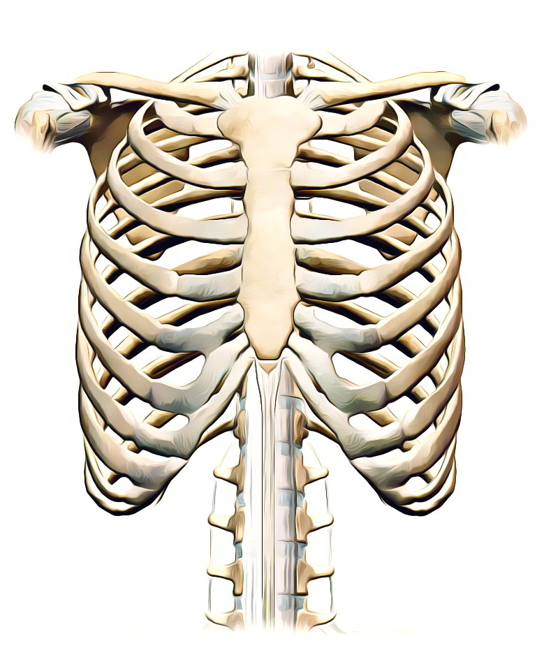
Rib Cage Anatomy : rib cage front | anatomia | Pinterest | Rib cage, Anatomy ... / From the anatomy of the human rib cage, we can tell that the human ribs bones have several parts:. Rib cage, in vertebrate anatomy, basketlike skeletal structure that forms the chest, or thorax, and is made up of the ribs and their corresponding attachments to the sternum (breastbone) and the vertebral column. Rib cage pain may be sharp, dull, or achy and felt at or below the chest or above the navel on either side. It is made up of 12 pairs of ribs. Each pair is numbered based on their attachment to the sternum, a bony process at the front of the rib cage which serves as an anchor point. 4 individual objects (spine portion, ribs, cartilages, sternum), sharing the same non overlapping uv layout map, material and pbr textures set.
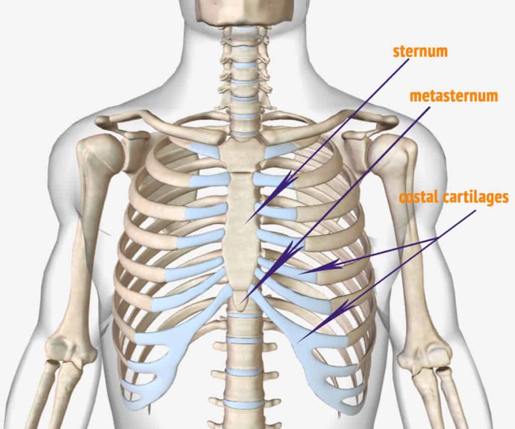
Two of the most notable organs behind the left side of the rib cage are the left lung and the spleen. The head only articulates with the body of the t1 vertebra and therefore only one articulatory surface is present. The number is the same for males and females. The ribs are a set of twelve paired bones which form the protective 'cage' of the thorax. They articulate with the vertebral column posteriorly, and terminate anteriorly as cartilage (known as costal cartilage).
Rib Cage Anatomy / The Thoracic Cage · Anatomy and ... from www.earthslab.com It consists of the 12 pairs of ribs with their costal cartilages and the sternum ( figure 7.32 ). Related posts of muscle anatomy rib cage muscle anatomy practice exam. Human rib cage anatomy 3d model. 5 out of 5 stars (81) $ 16.71. It may occur after an obvious injury or without explanation. From the anatomy of the human rib cage, we can tell that the human ribs bones have several parts: The lungs are responsible for processing oxygen through the body, while the spleen filters the blood and protects against some bacteria. The first rib is the widest, shortest and has the sharpest curve of all the ribs.
Rib cage, in vertebrate anatomy, basketlike skeletal structure that forms the chest, or thorax, and is made up of the ribs and their corresponding attachments to the sternum (breastbone) and the vertebral column.
We cover the different bones that make up the rib cage and some of the functions of. Muscle anatomy practice exam 12 photos of the muscle anatomy practice exam anatomy muscle practice questions, muscle anatomy practice exam, human muscles, anatomy muscle practice questions, muscle anatomy practice exam Rib cage pain can be caused. Related posts of rib cage diagram with organs abdominal cavity chart. Rib cage, in vertebrate anatomy, basketlike skeletal structure that forms the chest, or thorax, and is made up of the ribs and their corresponding attachments to the sternum (breastbone) and the vertebral column. The first rib is attached to thoracic vertebra. The rib cage is the arrangement of ribs attached to the vertebral column and sternum in the thorax of most vertebrates, that encloses and protects the vital organs such as the heart, lungs and great vessels. It may occur after an obvious injury or without explanation. In this video, we explore:1) the anatomy of the sternum2) the anatomy and differences between the three classes of ribs3) the anatomy and differences between. They articulate with the vertebral column posteriorly, and terminate anteriorly as cartilage (known as costal cartilage). The thoracic cage consists of the 12 thoracic vertebrae, the associated intervertebral discs, 12 pairs of ribs with their costal cartilages, and the sternum. The first rib is the widest, shortest and has the sharpest curve of all the ribs. The number is the same for males and females.
There are twelve (12) pairs of ribs and all articulate posteriorly with the thoracic vertebrae. In the front, ribs one to seven attach with cartilage to your sternum. Each pair is numbered based on their attachment to the sternum, a bony process at the front of the rib cage which serves as an anchor point. In this video we discuss the structure of the rib cage or thoracic cage. The head only articulates with the body of the t1 vertebra and therefore only one articulatory surface is present.
Antique Engraving Illustration Rib Cage Stock Vector Art ... from media.istockphoto.com The head only articulates with the body of the t1 vertebra and therefore only one articulatory surface is present. As part of the bony thorax, the ribs protect the internal thoracic organs. Anatomy the rib cage is a bony structure found in the chest (thoracic cavity). The thoracic cage (rib cage) forms the thorax (chest) portion of the body. As in the typical ribs, the tubercle has a facet for articulation with the transverse process of vertebrae. The cartilage strips are called costal cartilage (costal is the anatomical adjective that refers to the rib) and connect on their other end to the sternum. At the chest, many rib bones connect to the sternum via costal cartilage,. Humans usually have 24 ribs, in 12 pairs.
The number is the same for males and females.
The thoracic cage (rib cage) forms the thorax (chest) portion of the body. Heart in thorax, highlighting the valves. Abdominal cavity chart 14 photos of the abdominal cavity chart abdominal cavity cancer, abdominal cavity contains, abdominal cavity diagram picture, abdominal cavity pain, abdominal cavity quadrants, abdominal cavity regions, air in abdominal cavity, fluid buildup in abdominal cavity, stomach, abdominal cavity cancer. Muscle anatomy practice exam 12 photos of the muscle anatomy practice exam anatomy muscle practice questions, muscle anatomy practice exam, human muscles, anatomy muscle practice questions, muscle anatomy practice exam Related posts of muscle anatomy rib cage muscle anatomy practice exam. The lungs are two separate but connected organs located in the upper chest, covered by the rib cage. Each pair is numbered based on their attachment to the sternum, a bony process at the front of the rib cage which serves as an anchor point. The first rib is the widest, shortest and has the sharpest curve of all the ribs. Head (caput costae) neck (collum costae) body, corpus costae; Blend (2.80 with cycles / eevee. Two of the most notable organs behind the left side of the rib cage are the left lung and the spleen. The superior surface is unique in that it is marked by two grooves that allow. Rib cage pain can be caused.
The superior surface is unique in that it is marked by two grooves that allow. Humans usually have 24 ribs, in 12 pairs. The thoracic cage (rib cage) is the skeletal framework of the thoracic wall, which encloses the thoracic cavity. In normal development, a baby is born with 12 pairs of ribs. Anatomy the rib cage is a bony structure found in the chest (thoracic cavity).
Anatomy of The Human Ribs - With Full Gallery Pictures ... from dislocatedrib.org Introduction to the structure of the ribcage and ribs: As part of the bony thorax, the ribs protect the internal thoracic organs. And the front part of the rib. Rib cage anatomy watercolor print, rib cage anatomy watercolor art, chest bones anatomy, thorax anatomy art print as85 genefyart. Abdominal cavity chart 14 photos of the abdominal cavity chart abdominal cavity cancer, abdominal cavity contains, abdominal cavity diagram picture, abdominal cavity pain, abdominal cavity quadrants, abdominal cavity regions, air in abdominal cavity, fluid buildup in abdominal cavity, stomach, abdominal cavity cancer. The superior surface is unique in that it is marked by two grooves that allow. Anatomy, rib cage, my heart is a flower, rib cage art, medical art, gift for doctor, botany, anatomical art, book page art, book art ambicerebral. Rib cage pain can be caused.
They articulate with the vertebral column posteriorly, and terminate anteriorly as cartilage (known as costal cartilage).
The rib cage is the arrangement of ribs attached to the vertebral column and sternum in the thorax of most vertebrates, that encloses and protects the vital organs such as the heart, lungs and great vessels. Introduction to the structure of the ribcage and ribs: The lungs are responsible for processing oxygen through the body, while the spleen filters the blood and protects against some bacteria. 4 individual objects (spine portion, ribs, cartilages, sternum), sharing the same non overlapping uv layout map, material and pbr textures set. And more specifically, the rib cage is an egg with planes. All your ribs attach to your thoracic vertebrae — t1 to t12 — at the back of your upper body. 5 out of 5 stars (81) $ 16.71. They articulate with the vertebral column posteriorly, and terminate anteriorly as cartilage (known as costal cartilage). In this video, we explore:1) the anatomy of the sternum2) the anatomy and differences between the three classes of ribs3) the anatomy and differences between. Related posts of rib cage diagram with organs abdominal cavity chart. Blend (2.80 with cycles / eevee. The cartilage strips are called costal cartilage (costal is the anatomical adjective that refers to the rib) and connect on their other end to the sternum. Related posts of muscle anatomy rib cage muscle anatomy practice exam.